A 46-year-old female presented to the OPD with history of right upper quadrant pain and fever for 2 weeks. The following images were obtained in the right upper quadrant and the epigastric region.
What is the most probable diagnosis?
A. Liver simple cyst
B. Pyogenic liver abscess
C. Liver metastases
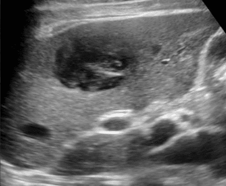
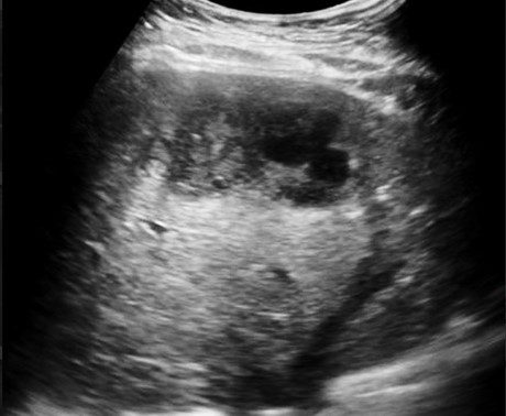
Image courtesy of UltrasoundCases.info owned by SonoSkills.
The most likely diagnosis is pyogenic liver abscess.
Explanation
Liver abscess may have variable appearance on ultrasound. Typical findings are a poorly demarcated lesions in the liver parenchyma which could be hypoechoic and have some hyperechoic areas. Hyperechoic gas bubbles may be seen sometimes. Color or power Doppler will show absence of flow in the middle of the lesion.
References


