
Do you know how to complete a focused cardiac ultrasound as an extension of the bedside exam? The LV should be 7cm in length and look like a football, a nicely shaped oval. If it...
Infographics
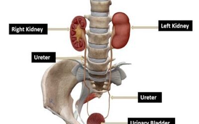
Renal POCUS
Looking for Evidence of Hydronephrosis Discover more POCUS applications through our POCUS Bytes Webinars.
Ultrasound Guided Venous Access Transverse Approach
This infographic explains the transverse approach, the views, needle placement and more. Discover more POCUS applications through our POCUS Bytes Webinars. Participate in Setting...
Lung Ultrasound
This infographic explores artifacts in point-of-care ultrasound (POCUS) to aid diagnosis when performing a ultrasound on the lungs. Note the differences between the A-lines and...
POCUS for Appendicitis
This infographic focuses on the best practices for using Point-of-Care-Ultrasound to accurately diagnose appendicitis. Note the differences in the scans showing longitudinal and...
RUSH Exam Protocol
How can point-of-care ultrasound help with rapid ultrasound for shock and hypertension? Use the nmenomic HIMAP-"ED" to help you remember. Heart IVC Morrison's Aorta...
Transverse Aortic Measurements
This infographic explores the use of point-of-care ultrasound to measure abdominal aortic aneurysms (AAA). Download the jpeg: Tips for Abdominal Aorta measurements Aortic...
Inferior Vena Cava (IVC) Exam
Do you know how to detect the fluid status during a point-of-care-ultrasound examination of the inferior vena cava (IVC)? Normal IVC Note the difference in diameter of the IVC...
POCUS Rush Protocol
This infographic provides information about the Rapid Ultrasound for Shock and Hypotension (RUSH) Exam Protocol.
POCUS DVT Assessment
This infographic provides information about point-of-care ultrasound and the 2-point compression DVT assessment.
Interested in certifying your entire team?
Connect with us and we'll prepare a custom package to meet your group's training and certification needs.
Recent Posts
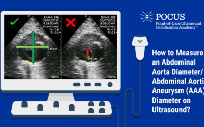
By Victor V Rao MBBS, DMRD, RDMS Introduction Abdominal aortic aneurysm (AAA) is defined as a focal dilatation of a segment of the abdominal aorta with an outer wall to...

By Shelby Jones Shelby is a senior at George Mason University studying public health. She is a current intern with POCUS Certification academy working within the...
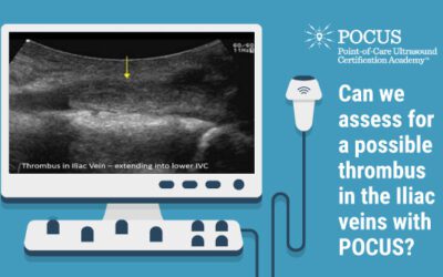
By Victor Rao MBBS, DMRD, RDMS - Abdomen, Ob/Gyn (APCA) Introduction Deep vein thrombosis (DVT) is a medical condition in which a thrombus develops in a deep vein,...
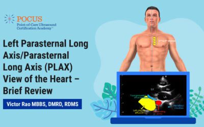
By Victor Rao MBBS, DMRD, RDMS - Abdomen, Ob/Gyn (APCA) Introduction The parasternal long axis (PLAX) view of the heart is a relatively easy view of the heart to obtain...

Remembering Pioneers in Radiology As Black History Month approached this year, we found ourselves inspired to dive into the history of medical imaging and its...

The use of point-of-care ultrasound (POCUS) has steadily increased in both clinical and community settings in recent years. It is one of the few diagnostic methods that...
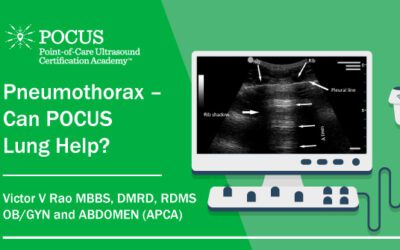
By Victor V Rao MBBS, DMRD, RDMS Ob/Gyn and Abdomen (APCA) Pneumothorax is the presence of air outside the lung and in the pleural space. The pleural space is the...
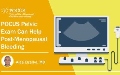
By Alaa Elzarka MD, PhD, OB GYN Lecturer, Alexandria Faculty of Medicine Abnormal uterine bleeding is considered a common problem for women during their childbearing...
By Victor Rao MBBS, DMRD, RDMS (APCA) Introduction Transabdominal ultrasound is a quick and easy modality to clinically assess a patient presenting with signs and...


